What Is Retinoblastoma?
Cancer starts when cells begin to grow out of control. Cells in nearly any part of the body can become cancer, and can spread to other areas. To learn more about how cancers start and spread, see What Is Cancer? For information about the differences between childhood cancers and adult cancers, see Cancer in Children.
Retinoblastoma is a cancer that starts in the retina, the very back part of the eye. It is the most common type of eye cancer in children. Rarely, children can have other kinds of eye cancer, such as medulloepithelioma, which is described briefly below, or ocular (eye) melanoma.
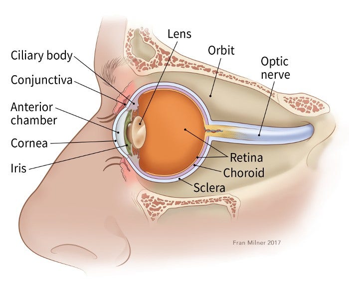
To understand retinoblastoma, it helps to know how the parts of the eye work.
The eye
The main part of the eye is the eyeball (also known as the globe), which is filled with a jelly-like material called vitreous humor. The front of the eyeball has a clear lens with an iris (the colored part of the eye that acts like a camera shutter), which allows light to enter the eye and focuses it on the retina.
The retina is the inner layer of cells in the back of the eye. It is made up of special nerve cells that are sensitive to light. These light-sensing cells are connected to the brain by the optic nerve, which runs out the back of the eyeball. The pattern of light (image) that reaches the retina is sent through the optic nerve to an area of the brain called the visual cortex, allowing us to see.
How does retinoblastoma develop?
The eyes start to develop well before birth. During the early stages of development, the eyes have cells called retinoblasts, which multiply to make new cells that fill the retina. At a certain point, these cells stop multiplying and become mature retinal cells.
Rarely, something goes wrong with this process. Instead of maturing, some retinoblasts continue to grow out of control, forming a cancer known as retinoblastoma.
The chain of events inside cells that leads to retinoblastoma is complex, but it almost always starts with a change (mutation) in the RB1 gene. The normal RB1 gene helps keep cells from growing out of control, but a change in the gene stops it from working like it should. Depending on when and where the change in the RB1 gene occurs, it can result in 2 different types of retinoblastoma.
Congenital (heritable) retinoblastoma
In about 1 out of 3 children with retinoblastoma, the abnormality in the RB1 gene is congenital (present at birth) and is in all the cells of the body, including all of the cells of both retinas. This is known as a germline mutation.
Despite this sometimes being called 'heritable' (or 'hereditary'), in most of these children, there is no family history of this cancer, and the RB1 gene change is not inherited from a parent. In these children, the gene change first occurs during early development in the womb. Only a small portion of the children born with this gene change inherit it from a parent.
Children born with a mutation in the RB1 gene usually develop retinoblastoma in both eyes (known as bilateral retinoblastoma), and there are often several tumors within the eye (known as multifocal retinoblastoma).
Because all of the cells in the body have the changed RB1 gene, these children also have a higher risk of developing cancers in other parts of the body.
- A small number of children with this form of retinoblastoma will develop another tumor in the brain, usually in the pineal gland at the base of the brain (a pineoblastoma). This is also known as trilateral retinoblastoma.
- For survivors of hereditary retinoblastoma, the risk of developing other cancers later in life is also higher than average (to learn more, see After Treatment for Retinoblastoma).
Sporadic (non-heritable) retinoblastoma
In about 2 out of 3 children with retinoblastoma, the abnormality in the RB1 gene develops in only one cell in one eye. It is not known what causes this change. A child who has sporadic (non-heritable) retinoblastoma develops only one tumor in one eye. This type of retinoblastoma is often found when the child is slightly older compared with those who have the heritable form.
Children with this type of retinoblastoma do not have the same increased risk of other cancers as children with congenital retinoblastoma.
For more on the heritable and non-heritable forms of retinoblastoma, see What Causes Retinoblastoma?
How does retinoblastoma grow and spread?
If retinoblastoma tumors are not treated, they can grow and fill much of the eyeball. Cells might break away from the main tumor on the retina and reach other parts of the eye, where they can form more tumors. These tumors might block the channels that let fluid circulate within the eye, raising the pressure inside the eye. This can cause glaucoma, which can lead to pain and loss of vision in the affected eye.
Most retinoblastomas are found and treated before they have spread outside the eyeball. But if they are not, retinoblastoma cells can spread to other parts of the body. The cells sometimes grow along the optic nerve and reach the brain. Retinoblastoma cells can also grow through the covering layers of the eyeball and into the eye socket, eyelids, and nearby tissues. Once the cancer is outside the eyeball, it can then spread to lymph nodes (small bean-sized collections of immune system cells) and to other organs such as the liver, bones, and bone marrow (the soft, inner part of many bones).
Intraocular medulloepithelioma
Medulloepithelioma is a very rare type of tumor that can start in the eye. It is not a type of retinoblastoma, but it's mentioned here because it also usually occurs in young children.
Medulloepitheliomas start in the ciliary body, which is near the front of the eye (see image above). Most of these tumors are malignant (cancerous), but they rarely spread outside the eye. They usually cause eye pain and loss of vision.
The diagnosis is made when a doctor finds a tumor in the eye by using an ophthalmoscope (an instrument that helps doctors to look inside the eye). As with retinoblastoma, the diagnosis is usually made based on where the tumor is inside the eye and how it looks. A biopsy (removing cells from the tumor to be looked at under a microscope) to confirm the diagnosis is almost never done because it might harm the eye or risk spreading the cancer outside of the eye.
Treatment for medulloepithelioma is almost always surgery to remove the eye. This usually gets rid of all of the cancer, as long as it was still only in the eye.
- Written by
- References
The American Cancer Society medical and editorial content team
Our team is made up of doctors and oncology certified nurses with deep knowledge of cancer care as well as journalists, editors, and translators with extensive experience in medical writing.
Hurwitz RL, Shields CL, Shields JA, et al. Chapter 27: Retinoblastoma. In: Pizzo PA, Poplack DG, eds. Principles and Practice of Pediatric Oncology. 7th ed. Philadelphia, Pa: Lippincott Williams & Wilkins; 2016.
Kaufman PL, Kim J, Berry JL. Retinoblastoma: Clinical presentation, evaluation, and diagnosis. UpToDate. Accessed at www.uptodate.com/contents/retinoblastoma-clinical-presentation-evaluation-and-diagnosis on September 18, 2018.
National Cancer Institute. Retinoblastoma Treatment (PDQ®). 2018. Accessed at www.cancer.gov/types/retinoblastoma/hp/retinoblastoma-treatment-pdq on September 18, 2018.
Rodriguez-Galindo C, Orbach DB, VanderVeen D. Retinoblastoma. Pediatr Clin North Am. 2015;62(1):201-223.
Last Revised: December 3, 2018
American Cancer Society medical information is copyrighted material. For reprint requests, please see our Content Usage Policy.
American Cancer Society Emails
Sign up to stay up-to-date with news, valuable information, and ways to get involved with the American Cancer Society.
Help us end cancer as we know it, for everyone.
![]()